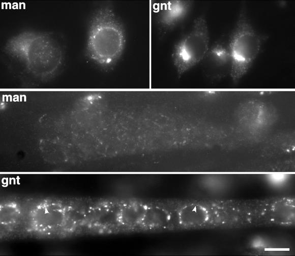
Figure 4.
PLA2 inhibitor ONO redistributes GC proteins to the ER in both myoblasts and myotubes. The culture medium of C2 myoblasts (top row) or myotubes (bottom two rows) was replaced with serum-free medium supplemented with 15 μM ONO. After 2 h, cultures were stained with anti-mannosidase (man) or anti-giantin (gnt) and viewed by conventional fluorescence microscopy. The pattern shows a redistribution of mannosidase and giantin to the ER (compare with Figure 1 as control). In myotubes, ONO causes a decrease in the fluorescence intensity of both markers, especially noticeable with mannosidase; giantin staining appears more fuzzy and punctate than in control myotubes with a fine continuous staining detectable around some nuclei (arrowheads). Bar, 10 μm.

