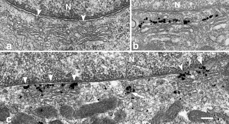
Figure 7.
ER-GC area around the nuclei of myotubes is labeled with anti-Sec13p in immunogold EM. After 2 d in fusion medium, C2 cultures were fixed with 2% glutaraldehyde, processed, and embedded for EM (a); other cultures were fixed with a mixture of acrolein-p-formaldehyde (b and c), and immunolabeled with mouse anti-GM130 (b) or rabbit anti-Sec13p (c) followed by a gold-conjugated secondary antibody and silver enhancement (see MATERIALS AND METHODS). In the microscope, we focused on the perinuclear area, which is easily identifiable. Each panel shows small Golgi stacks next to a nucleus (N). Notice in a the evaginations of the outer nuclear membrane (arrowheads) pointing toward the GC, and the electron-dense coat associated with them, which resembles the COPII coat (Barlowe, 1998). (b) Position of the cis-GC labeled with anti-GM130. (c) Discrete aggregates of Sec13p covering the outer nuclear membrane and the adjacent area facing the cis-GC (arrowheads). Not every aggregate is asssociated with the nuclear membrane (arrow). Bar, 200 nm.

