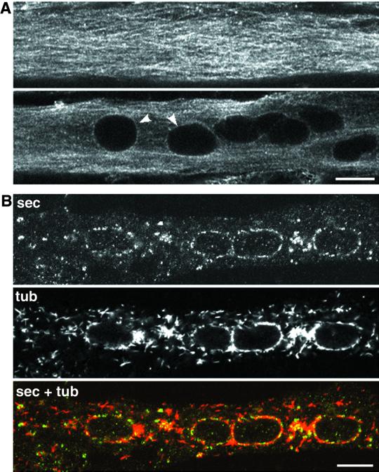
Figure 8.
ER exit sites are close to microtubule minus ends in myotubes. Microtubules in untreated myotubes (A) and in myotubes in which microtubules had been left to recover for 5 min after depolymerization (B) were stained with anti-tubulin and observed in the confocal microscope. Close to the cell surface (A, top), there is a dense network of microtubules without clear nucleation points. In the core of the myotube (A, bottom), there is a perinuclear ring of tubulin (arrowheads) in addition to mostly longitudinal microtubules. (B) C2 myotubes were placed on ice for 10 min, and then warmed up at 37°C in nocodazole for 1 h to depolymerize the microtubules. Nocodazole was then washed out. After a 5-min recovery, cultures were fixed and stained with anti-tubulin (tub) and anti-Sec31p (sec). The microtubules had already reformed a perinuclear belt and had regrown from many cytoplasmic sites as well. Most ER exit sites (labeled with anti-Sec31p) are in proximity of a site of microtubule nucleation, which corresponds to the minus end of the microtubules. Bar, 10 μm.

