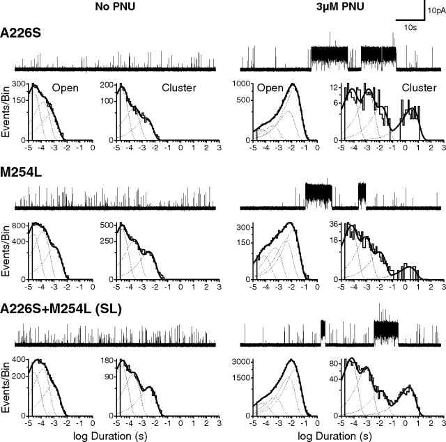
Figure 3.
Mutations that markedly reduce PNU potentiation at the whole-cell level exhibit potentiation at the single-channel level. Current traces with associated open time and cluster duration histograms from mutant α7 receptors (A226S; M254L; A226S+M254L double mutant), both in the absence (left) and presence (right) of 3 μm PNU in the pipette solution, are shown. Openings were elicited by 100 μm ACh; all recordings were made in the cell-attached patch configuration with an applied voltage of −70 mV. Single-channel traces are shown at a bandwidth of 5 kHz, and openings are upward deflections.

