
Figure 7.
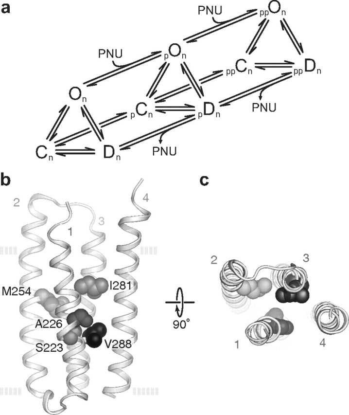
a, A possible mechanism for PNU potentiation. The scheme does not include agonist binding steps and assumes closed (C), open (O), and desensitized (D) states are bound with an optimal number of n agonists. The scheme also includes a minimum of two classes of PNU potentiation (p, pp) thought to relate to different levels of PNU occupancy. b, c, Structural determinants of PNU potentiation. Homology model (Cheng et al., 2006) based on Protein Data Bank identifier 2BG9 (Unwin, 2005) of the isolated transmembrane region of a single α7 subunit as viewed from the plane of the membrane (b) and the synaptic space (c). The side chains of the five amino acids mutated in the quintuple (TSLMF) mutant are shown as space-filling spheres. The four transmembrane helices (TM1-4) are also labeled. The dashed lines in b represent the approximate location of the plasma membrane.