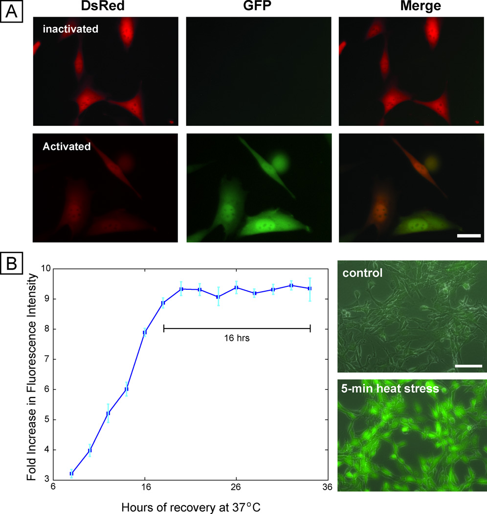
Fig. 1. Cell-based stress sensor.
(A) Fluorescence microscopy images of cells before and after stress activation using sodium arsenite, showing constitutive expression of DsRed and inducible expression of EGFP. Scale bar 25 µm. (B) Dynamics of expression of sensor after removal of sodium arsenite stressor, showing stable EGFP expression for 16 hours (left panel). Right panel shows the response of the sensor to short duration (5 minute) heat stress, with robust expression of EGFP.

