
Figure 6. PDF outputs are affected upon acute silencing.
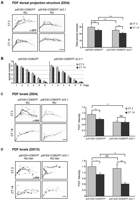
(A-B) The complexity of PDF axonal arborisations is compromised during the silenced phase. (A) Left panel. Representative confocal images of fly brains stained for GFP taken at the early subjective day (CT2) and early subjective night (CT14) on DD4. Right panel. Total number of axonal crosses. Control pdf-GS>CD8GFP flies kept as adults on RU displayed circadian remodelling of the axonal terminals. pdf-GS>CD8GFP;kir2.1 flies displayed reduced complexity throughout the day, although circadian structural plasticity was still evident. Forty to fifty flies were analysed per group. (B) Number of intersections between each concentric ring and the axonal projections in control and electrically silenced flies. The complexity of the axonal arbors is consistently lower in the night-time conformation. (C-D) Analysis of PDF levels. Left panels. Magnified views of the LNvs projections in the dorsal region stained for PDF. Brains were fixed at CT 2 and CT 14. Samples were taken 4 days after the last light-dark transition (DD4, C) and 5 days after RU removal (DD13, D). Experiments were repeated at least 3 times and a minimum of 30 brains were analysed per timepoint. Right panels. Quantitation of the average intensity of the LNvs dorsal projections during the silenced (DD4, C) and recovered (DD13, D) phase was performed blind. Error bars represent the standard error of the mean. Statistical analysis for total axonal crosses and PDF levels included ANOVA followed by a pairwise comparison (Bonferroni test). *** indicates p<0,001, ** indicates p<0,01. NS: Non Significantly different. Scale bar: 10μm.