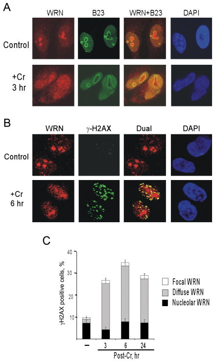
Figure 2. Cr(VI) causes dispersion of WRN from nucleolus.
(A) Confocal images of control and 20 μM Cr(VI)-treated NF cells immunostained for WRN and the nucleolar marker B23. (B) A representative confocal image of Cr-treated cells displaying colocalization WRN with γ-H2AX foci. (C) Presence of γ-H2AX foci in NF cells with different patterns of WRN staining. Cells were treated with 20μM Cr(VI) and fixed for immunostaining at indicated times. Data are means±SD from three slides with >100 cells scored per slide.

