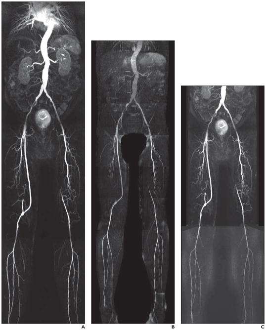
Fig. 1. 61-year-old man with diabetes.
A–C, Contrast-enhanced MR angiography (CEMRA) image (A), quiescent-interval single-shot unenhanced MR angiography image (B), and CEMRA image with superimposed time-resolved angiography with interleaved stochastic trajectories (C) show moderately diseased right external iliac and common femoral artery. Note right external iliac artery stent in situ. There is focal narrowing of left external iliac artery. Femoral-popliteal graft with three-vessel runoff. Occluded left superficial femoral artery with reconstitution above knee and three-vessel runoff.

