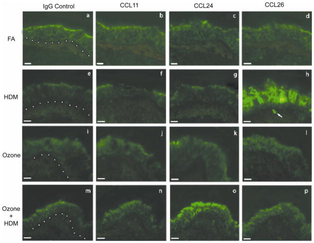
Figure 6. Effect of ozone and allergen exposure on CCL11, CCL24 and CCL26 protein expression in infant monkey airways.
Immunofluorescence staining for CCL11, CCL24 and CCL26 was conducted on adjacent cryosections obtained from a midlevel intrapulmonary airway of the left caudal lobe from each animal at 3 months of age. One representative infant monkey from each FA (a–d), HDM (e–h), ozone (i–l), and ozone + HDM (m–p) exposure group is shown. Purified goat IgG was used as a negative control (a, e, i, m). Each image panel contains a portion of the airway lumen (top), airway epithelium (center), and airway interstitium (bottom). The dotted line represents the basement membrane zone in panels a, e, i and m. The arrow in (h) points to a CCL26+ cell within the airway interstitium (scale bar= 10 μm).

