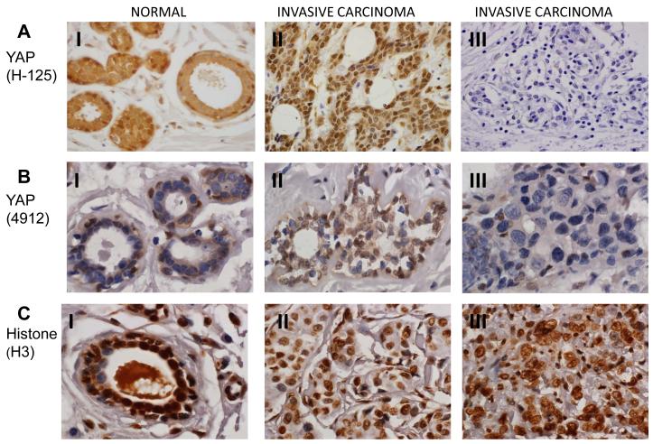
Fig. 1.
YAP immunostaining in normal and invasive carcinoma breast tissues. (60X magnification). A and B (I): Immunohistochemistry with two different YAP antibodies, H-125 (A) and 4912 (B) showing that expression of YAP is strongly positive with high level of nuclear staining in basal cells of normal breast ducts and in the cytoplasm of luminal cells. A and B (II): Representative invasive carcinoma tissues that are positive for YAP expression. A and B (III): Representative invasive carcinoma tissues that are negative for YAP expression. C (I): Immunstaining of normal breast tissue showing strong expression of histone H3. C (II): Immunostaining of histone H3 of invasive carcinoma tissues those are positive for YAP expression. C (III): Immunostaining of histone H3 of invasive carcinoma tissues those are negative for YAP expression.

