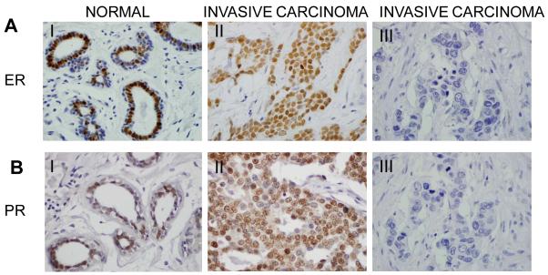
Fig. 2.
ER and PR immunostaining in normal and breast carcinoma. (60X magnification). A and B. Immunostaining was performed to detect the expression of ER (A) or PR (B) in normal and invasive carcinoma tissues. I): Strong positive and nuclear focal expression of ER (A) or PR (B) was observed in cells of normal ducts. II): Representative invasive breast carcinoma that is positive for ER (A) or PR (B) expression. III): Representative invasive breast carcinoma that is negative for ER (A) or PR (B) expression.

