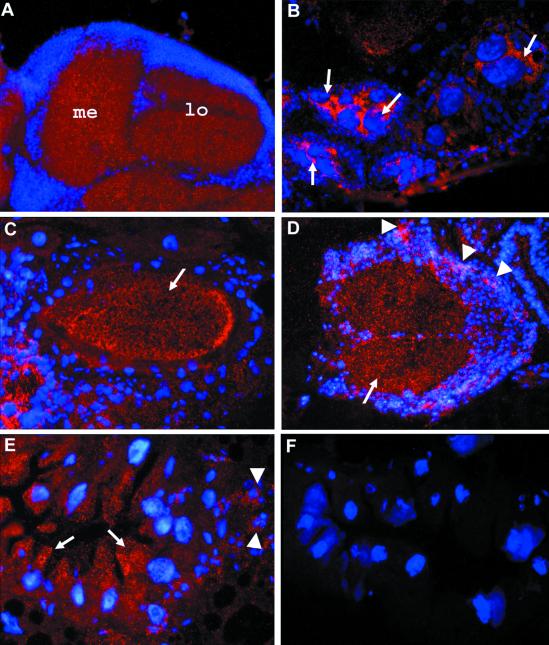
Figure 3.
Immunohistochemical localization of mtSSB. Cryosections of various tissues and stages (10 μm thick) were subjected to immunofluorescence analysis using a rabbit mtSSB antiserum. The tissue was counterstained with DAPI to visualize nuclei. (A) Horizontal section through the visual system of an adult head. Strong labeling is seen in the neuropil region. (B and C) Horizontal sections through the abdomen of an adult female. In B, a high level of mtSSB is seen in nurse cells (arrows). (C) The protein is transported into the oocyte (arrow). (D) A cross-section through a third instar larval ventral ganglion shows expression of mtSSB in the neuropil region (arrow) and elevated expression in proliferating neuroblasts (arrowheads). (E) Cross-section through the midgut of a third instar larvae. A high concentration of mtSSB is found in the apical region of epithelial cells facing the gut lumen (arrows) and in islets of small imaginal cells, giving rise to the adult midgut tissue (arrowheads). (F) Comparable section as in E from a homozygous lopo1 larva. No specific staining is seen.

