
Fig. 6. Detection of stromal cell-derived factor-1 (SDF-1) expression in the major pelvic ganglion. Rats (n = 5 in each group) were treated with sham operation or bilateral cavernous-nerve (CN) crush, and then intracavernously injected with 5-ethynyl-2-deoxyuridine (EdU)-labeled adipose-derived stem cells (ADSC). One day later, both EdU and SDF-1 were clearly visible in the major pelvic ganglia (MPG) of the (a) Crush+ADSC group, but not of the (b) Sham+ADSC group. (c) Higher magnification of the boxed area in panel A further shows the close association between SDF-1 expression and ADSC. Sections were stained for EdU, SDF-1, and 4′,6-diamidino-2-phenylindole (DAPI). Original magnification: ×100. (d) The graph demonstrates the expression levels of SDF-1 in the MPG 1 d after CN sham or crush injury.
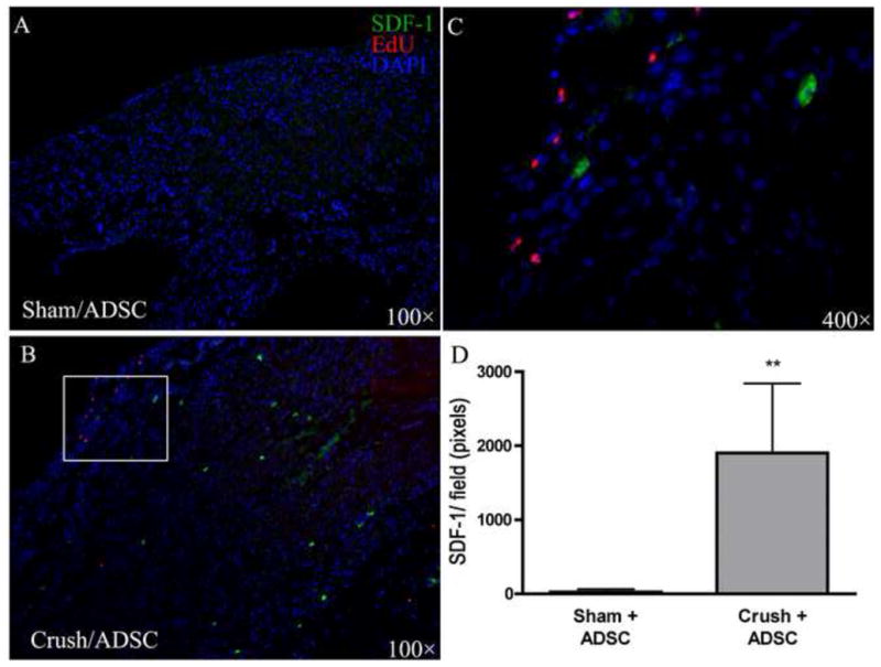
**p < 0.0001 versus Sham+ADSC.