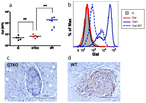
Figure 1. Islets from galactosyl transferase-hemizygous neonatal porcine donors express Gal.
(a) Islet transplant preparations isolated from WT neonatal donors had significantly higher Gal expression as determined by flow cytometric analysis than GTKO counterparts (p<0.008, n=5); GTKO islet Gal expression was not significantly different than WT islets stained with an isotype control. One WT islet preparation had an MFI of Gal expression that was significantly lower than other WT preparations (denoted with†). (b) Histogram demonstrating Gal expression phenotype for islet preparations from representative GTKO and WT donors compared to an isotype control. One WT islet preparation demonstrated an intermediate Gal expression phenotype (represented with dashed line). (c,d) Immunohistochemical analysis of transplanted intrahepatic islets demonstrates presence of Gal epitope (brown stain) on WT (d) but not GTKO (c) islets. P-value determined using Mann-Whitney test. **p <0.01; MFI – mean fluorescence intensity; IC – isotype control; Gal-INT† – intermediate Gal expression phenotype.

