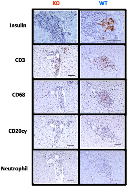
Figure 6. Gal-deficient islets are not protected from eventual cellular rejection.
Immunohistochemical analysis of intrahepatic islets from recipients experiencing functional xenograft rejection demonstrated dense lymphocytic infiltrates with loss of insulin positivity. This infiltrate stained heavily for CD3+ T cells, and moderately for CD68+ macrophages. CD20+ B cells and neutrophils did not appear to comprise a significant portion of the infiltrating cells.

