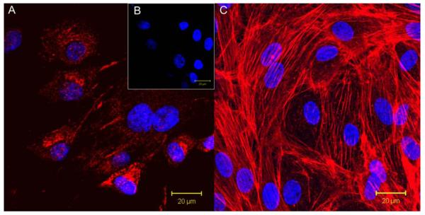
Figure 7.
(A) BMEOCs stained for vWF (red) with a DAPI (blue) nuclear stain. (B) Control for vWF stain with the DAPI nuclear stain. (C) Endothelial cell monolayer on 15 wt% prTE after 48 hours in culture. Nuclei are stained with DAPI and the cytoskeleton (f-actin) is stained with rhodamine phalloidin (red). BMEOCs attached and spread on the prTE scaffold.

