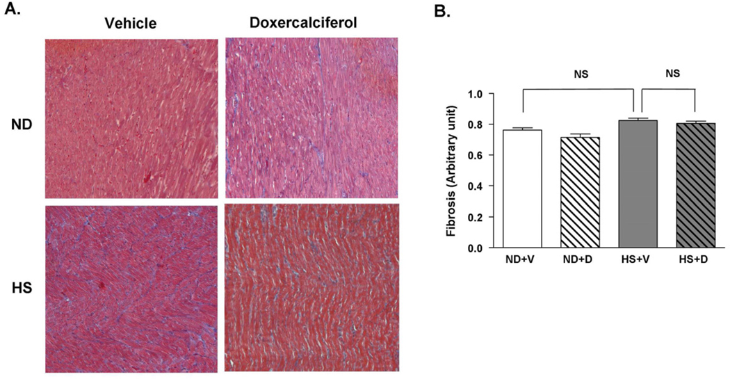
Figure 5.
Histological analysis of cardiac fibrosis in DSS rats with or without doxercalciferol. (A) Representative Masson’s trichrome staining of the heart sections. (B) There was no significant difference in cardiac fibrosis in all experimental groups. 25–30 fields at ×10 objective per animal were measured. Fields with >5% fibrosis area were counted and divided by the total number of fields observed. ND=normal diet, HS=high salt diet, V=vehicle, D=doxercalciferol *p<0.05, N=6–9.

