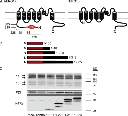
Figure 1.
hERG1a NTRs form polypeptides. (A) Schematic of hERG1a channel subunit and hERG1b subunit. Arrows indicate positions of truncation mutants in the hERG1a NTR. (B) Schematic of hERG1a NTR polypeptides formed from truncation mutants. (C) Western blot of lysates from hERG1a/hERG1b stable cell line transfected with hERG1a NTR plasmids. (Top) Bands corresponding to hERG1a and hERG1b; (middle) bands corresponding to PDI, which is used as a loading control; and (bottom) bands corresponding to hERG1a NTR polypeptides as indicated. Lanes corresponding to control (Kir2.1) and mock (transfection reagent) experiments were also included, as indicated.

