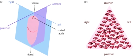
Figure 1.
(a) Diagram of the mouse embryo 8 days post-fertilization, showing with dorsal–ventral, anterior–posterior and left–right axes, the ventral node structure being indicated. Adapted from Nonaka et al. (2002). (b) Schematic of the node, containing an approximately triangular array of tilted, clockwise rotating cilia. The tilt results in the cilia paths appearing elliptical when viewed from above. The black lines depict instantaneous positions of cilia, and the ellipses and arrows depict the path of rotation.

