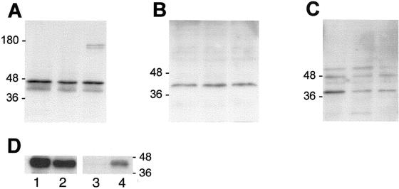
Figure 2.
Western blot analysis of Cx43, Cx40, and Cx37 expression. (A–C) Aliquots of protein extracts collected from parental bEnd.3 cells (left lanes), bEnd.3/3243H7 cells (middle lanes), and bEnd.3/Cx43βGal cells (right lanes) were subjected to SDS/PAGE in 12% polyacrylamide gels. After transfer to nitrocellulose membranes, the blots were probed using Cx43 (A), Cx40 (B), or Cx37 (C) antibodies, with detection performed by chemiluminescence. (A) Anti-Cx43 antibody revealed several bands at 41–47 kDa, corresponding to the endogenous Cx43 protein and/or the chimeric 3243H7 protein, as well as two higher molecular weight bands at ∼160 kDa, corresponding to the Cx43βGal fusion protein. (B) Anti-Cx40 antibody revealed a band at 42 kDa, corresponding to the Cx40 protein. (C) Anti-Cx37 antibody revealed a band at 39 kDa, corresponding to the Cx37 protein, as well as two nonspecific bands at ∼48–60 kDa, which were also observed after incubation with preimmune serum. (D) Protein extracts of parental bEnd.3 cells (lanes 1 and 3) and bEnd.3/3243H7 cells (lanes 2 and 4) were immunoprecipitated with anti-HA antibodies. Supernatants (lanes 1 and 2) and immunoprecipitates (lanes 3 and 4) were collected, blotted, and probed with anti-Cx43 antibodies. Native Cx43 was slightly less abundant in bEnd.3/3243H7 cells (lane 2) than in parental cells (lane 1). The precipitated chimeric connexin could be specifically recognized in bEnd.3/3243H7 cells (lane 4).

