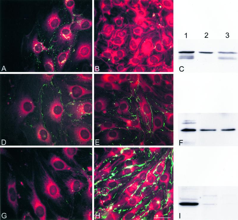
Figure 5.
Cx43 is up-regulated and Cx37 down-regulated at the wound edge in parental bEnd.3 cells, 24 h after wounding. (A, B, D, E, G, and H) Representative fluorescence micrographs of the wound (A, D, and G) and outside the wound regions (B, E, and H) of parental bEnd.3 cells after immunostaining with anti-Cx43 (A and B), anti-Cx40 (D and E), or anti-Cx37 (G and H) antibodies. Cells are counterstained with Evans Blue. (A) Punctate Cx43 immunoreactivity (green labeling) was induced along the lateral borders of adjacent cells in the wound area. (B) This immunoreactivity was virtually absent at cell-to-cell contacts in outside the wound regions. (D and E) Cx40 immunoreactivity can be detected in regions of cell-to-cell contact in both wound and outside of the wound regions. (G) Cx37 immunoreactivity was not detected in the first rows of cells lining the wound. (H) In contrast, this immunoreactivity is abundantly detected in areas outside the wound. Bar, 30 μm. (C, F, and I) Western blots of proteins extracted from HeLa transfectants (lanes 1), confluent (lanes 2), or multiple wounded cultures of bEnd.3 cells (lanes 3) probed with Cx43 (C), Cx40 (F), or Cx37 (I) antibodies. (C) Anti-Cx43 antibody revealed several bands at 41–47 kDa in control HeLaCx43 cells. A similar band pattern was detected in bEnd.3 cells from multiple wounded cultures. In confluent bEnd.3 cell cultures only a single band a 47 kDa was detected. (F) Anti-Cx40 antibody revealed a major band at 42 kDa in control HeLaCx40 cells. Cx40 bands of equal intensity were detected in multiple wounded and confluent bEnd.3 cell cultures. (I) Anti-Cx37 antibody revealed a major band at 39 kDa in control HeLaCx37 cells. This band could be readily detected in confluent bEnd.3 cell cultures, whereas it was virtually absent in multiple wounded cultures.

