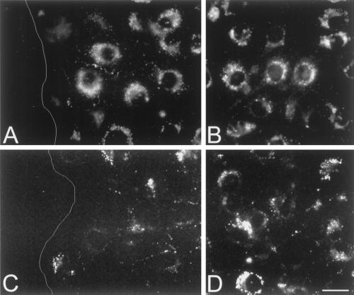
Figure 7.
Inhibition of wound-induced Cx43 up-regulation in bEnd.3 cells expressing dominant negative connexins. (A–D) Representative fluorescence micrographs of the wound (A and C) and outside the wound region (B and D) of bEnd.3/3243H7 cells (A and B) and bEnd.3/Cx43βGal cells (C and D), 24 h after wounding. Cx43 immunoreactivity was detected predominantly in the perinuclear region. No obvious increase in Cx43 labeling was observed in the wound region of either transfected clone. Bar, 22 μm. White line (A and C) represents the wound edge.

