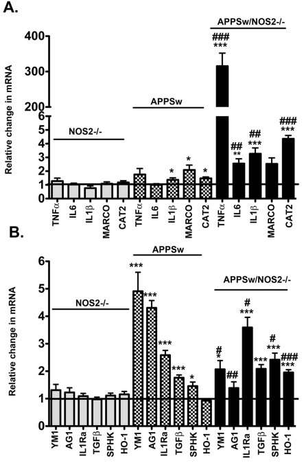
Figure 1. Comparison of mRNA expression levels for classical activation, alternative activation and acquired deactivation genes associated with the brain's innate immune response between a mouse model of amyloid deposition and a model of disease progression.
Results represent the means±S.E.M. fold-change in mRNA levels for classical activation genes (A) and for alternative activation genes and acquired deactivation genes (B) in 52–60-week-old NOS2−/−, APPSw and APPSw/NOS2−/− mice brain samples compared with WT control mice of the same age (n = 5–7 mice per strain). *P<0.05, **P<0.01, ***P<0.001 compared with the NOS2−/− control mice in each case. #P<0.05, ##P<0.01, ###</emph>P<0.001 compared with the parent strain.

