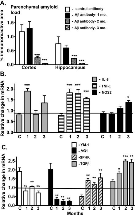
Figure 5. Passive immunization alters the immune profile in APPSw mice.
(A) Parenchymal amyloid load in the cortex and hippocampus. As described in the Materials and methods section, 19-month-old APPSw mice were assigned to one of four groups, control antibody for 3 months or anti-Aβ antibody 2286 (Rinat Neurosciences) for 1, 2 or 3 months (n = 4/group). The start of dosing was staggered such that all mice were of the same age when killed. The level of amyloid immunostain in brains from passively immunized APPSw mice was measured by image analysis of immunostained regions as described in the Materials and methods section. Results are presented as the means±S.E.M. of the % immunoreactive area. (B, C) Results represent the means±S.E.M. fold-change in mRNA levels for selected classical activation genes (B) and for alternative activation and acquired deactivation genes (C) in immunized compared with control mice starting at 9 months of age (n = 4 mice/group). *P<0.05, **P<0.01, ***P<0.001 compared with control mice.

