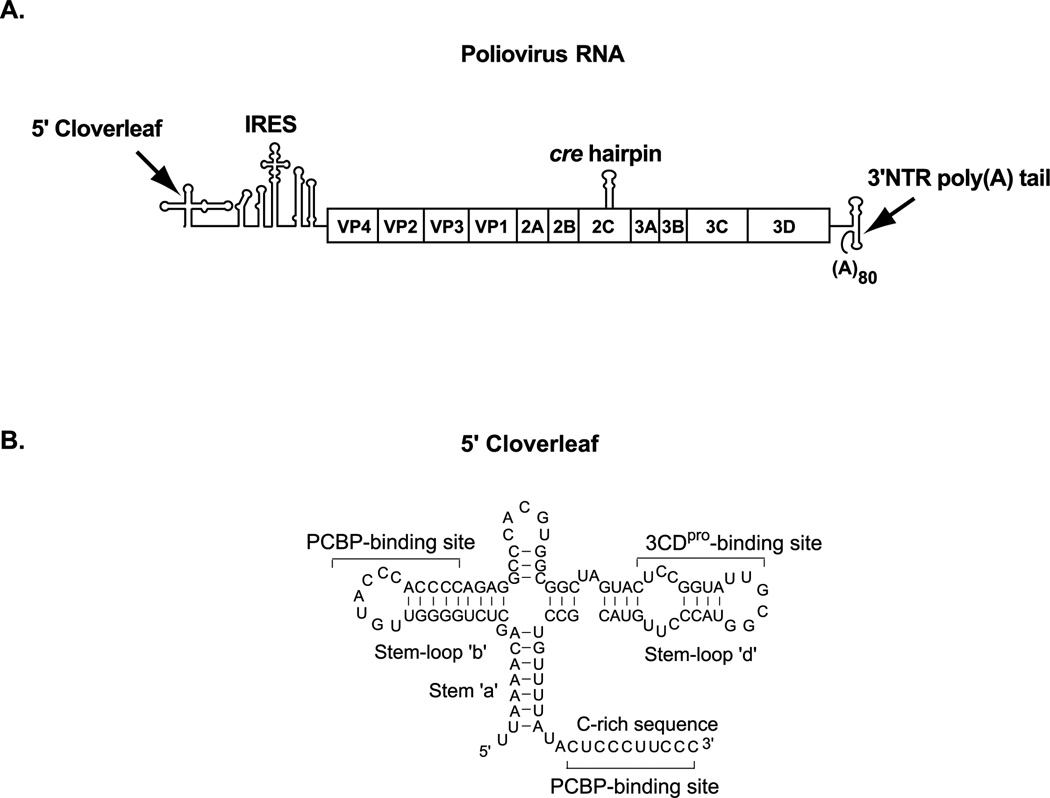
Figure 1.
(A). A schematic diagram showing the RNA structures in the poliovirus RNA genome which perform non-templated RNA functions during viral replication. The 5’ cloverleaf (5’CL) and the internal ribosome entry site (IRES) are present in the 5’ non-translated region (NTR) of the genome. The open reading frame in the viral genome encodes a polyprotein which is cleaved to generate the structural and replication proteins. The cre hairpin is present in the 2C coding region and the 3’ NTR and poly(A) tail is at 3’ end of the genome. (B). The secondary structure and nucleotide sequence of the 5’CL showing the binding sites for the cellular poly(C) binding protein (PCBP) and viral protein, 3CDpro.

