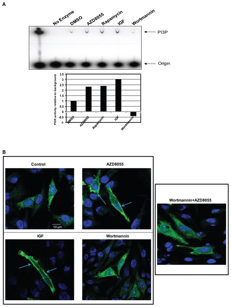
Figure 3. kinase inhibition leads to PI3K activation.
(A) MCF-7 cells were treated with 500nM of AZD8055, 50nM of rapamycin, or 100nM of wortmannin for four hours; or 60 ng/mL of IGF for ten minutes and a PI3K activity assay was performed. The product, PI3P, was resolved by thin layer chromatography and detected by autoradiography. A lane with a purified PI3K enzyme was used as a positive control and a lane without enzyme was used as a negative control for the assay. The results were quantified by densitometry (lower panel). (B) The pcDNA3-AKT-PH-GFP vector was transfected into HeLa cells. Twenty-four hours after the transfection, the cells were treated with either DMSO, 500nM of AZD8055, 100nM of wortmannin or combination of wortmannin and AZD8055 (wortmannin was added thirty minutes prior to AZD8055 for the combination treatment) for four hours or with 60 ng/mL of IGF for ten minutes. The GFP signal was detected using confocal microscopy. Shown are representative cells.

