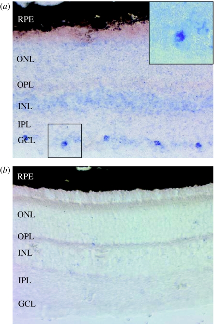
Figure 3.
Tissue in situ hybridization. Adult dunnart retinal sections probed with (a) Opn4 antisense riboprobe and (b) sense control riboprobe. Opn4 shows staining restricted to a subset of cells in the ganglion cell layer. No signal is seen with the sense probe. Inset in (a) is a magnification of the highlighted area. GCL, ganglion cell layer; IPL, inner plexiform layer; INL, inner nuclear layer; OPL, outer plexiform layer; ONL, outer nuclear layer; RPE, retinal pigmented epithelium.

