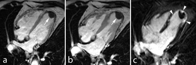
Fig. 10.
Old anterior MI in 75 year old man presenting with heart failure symptoms. Horizontal long–axis cine MRI (a) and early (b) ce–MRI show apical aneurysm with large typically hypoenhanced thrombus (arrows, a,b). Late ce–MRI in the same plane shows apical transmural enhancement (arrowheads, c).

