
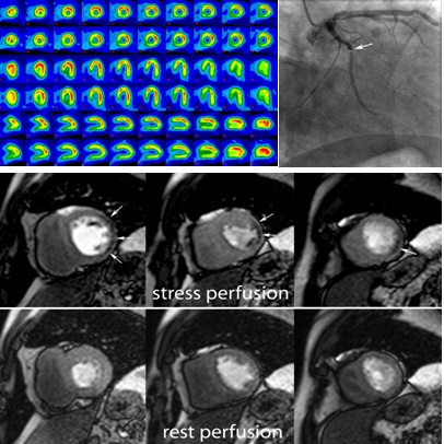
Fig. 5MRI study (rest and stress perfusion) in a 52 year old male with stable angina. Rest and stress (*) SPECT were negative, showing no induced perfusion defects (a). Midventricular short-axis cardiac MRI images (b) of first pass perfusion during rest (bottom) show no perfusion defects while during dipyridamole vasodilatatory stress (top) show a subendocardial perfusion defect in the lateral wall (arrows). Coronary angiography (c) confirmed a significant (90%) stenosis of the first lateral branch of Cx (arrow, vessel larger than the Cx).