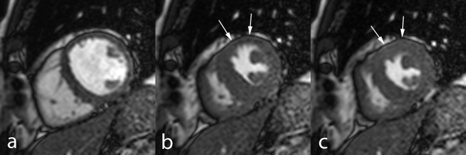
Fig. 6.
MRI study (dobutamine–atropine stress) in 60–year–old man. Midventricular cardiac short–axis cine MRI at end–diastole (a), end–systole (b), and isovolumic relaxation (c). At maximal stress, during systole (b), the ischemic myocardium in the anterior wall is akinetic (arrows). However, during isovolumic relaxation (c) while the non–ischemic regions start to relax, myocardial thickening can be is seen in the anterior LV wall (arrows), i.e. post–systolic contraction of the ischemic myocardium. Coronary angiography showed significant stenosis in the proximal LAD coronary artery.

