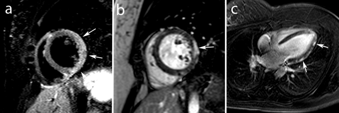
Fig. 7.
MRI study in an 18 year old male with acute chest pain in which acute myocarditis was diagnosed. Mid ventricular short axis T2–weighted STIR image shows edema (increased signal intensities) in the lateral wall (arrows, a) and short and vertical long–axis ce–MRI show typical subepicardial enhancement in the lateral wall (arrows, b,c).

