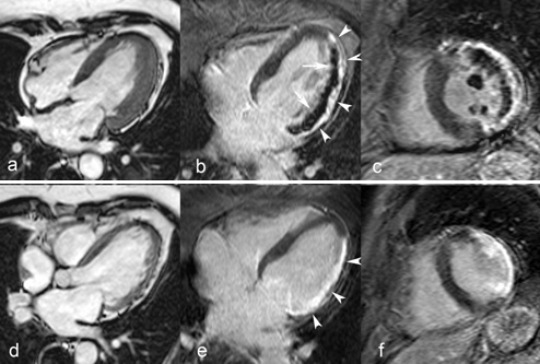
Fig. 8Extensive lateral MI in 68 year–old man studied 1 week and 4 months after the acute event. Proximal Cx coronary artery occlusion treated by primary PCI but complicated by no reflow. Cine MRI in horizontal long–axis shows thickened LV lateral wall due to myocardial edema and inflammation in the acute phase (a), followed by marked wall thinning at 4 months (d). Horizontal long–axis and midventricular short–axis late ce–MRI show an extensive subendocardial no reflow zone at 1 week (arrows b,c) and transmural enhancement of the lateral wall (arrowheads, b, c, e, f). Both papillary muscles are involved (no reflow at 1 week and enhancement at 4 months). At follow–up strong, homogeneous enhancement of the scarred myocardium (rrowheads, e).
