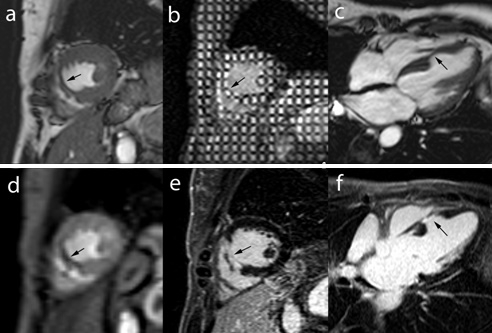
Fig. 9MRI study in a 47 year–old female with LV septal wall motion abnormalities (incidental findings) on echocardiography probably due to an embolic occlusion of a septal LAD coronary artery branch. Midventricular short–axis and horizontal long–axis cine MRI (a,c) and short–axis tagging MRI (b) show marked mid septal wall thinning with dyskinetic wall motion (arrows). First–pass perfusion MRI (d) and late ce–MRI (e,f) show a perfusion defect (d) and transmural enhancement (e, f) within the thinned myocardium (arrows).
