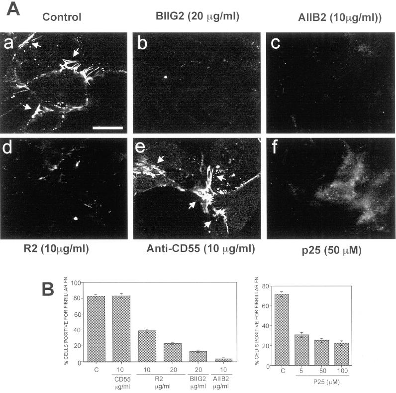
Figure 2.
The effect of disruption of uPAR-integrin association on FN fibrillogenesis. (A) LK25 cells plated on gelatin (10 μg/ml)-coated coverslips for 60 min were preincubated with medium alone (a) or with antibodies to α5β1-integrins (BIIG2 20 μg/ml, b), β1-integrin (AIIB2 10 μg/ml, c), uPAR (epitope in domain-III, R2, 10 μg/ml, d), CD55 (CD55/DAF, 15 μg/ml, e), or medium with 50 μM peptide p25 (f), for 20 min (AIIB2 and BIIG2 are function-blocking antibodies). Human FN (5 μg/ml) was then added to all wells, and the cells were incubated overnight to allow for FN fibrillogenesis. Cells were examined by IF confocal laser scanning microscopy. The arrows indicate FN fibrils between or beyond cell bodies. (B) Quantitation of FN fibril-positive cells. At least 200 cells were scored per treatment in duplicate. Treatments in the left graph are: C, control medium; CD55, antibody anti-CD55 (10 μg/ml); R2, uPAR antibody (10 and 20 μg/ml); BIIG2, anti-α5β1 antibody (20 μg/ml); AIIB2, anti-β1 antibody (10 μg/ml). Treatments in right graph are: P25, peptide 25 that disrupts uPAR-integrin interaction (5, 50, and 100 μM). Quantitation of FN fibril-positive cells was performed as described in Figure 1, C and D, using DAPI staining. Bar, 40 μm

