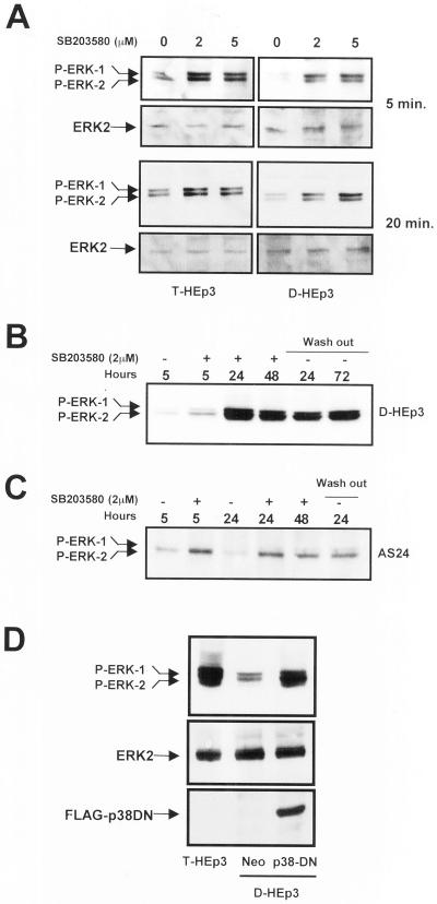
Figure 4.
Analysis of cross-talk between ERK and p38 pathways. (A) Effect of p38 inhibition on ERK activation (short-term treatment). Serum-starved T-HEp3 (left) or D-HEp3 (right) cells were treated for 5 or 20 min with 0, 2, and 5 μM SB203580 in DMSO, then lysed, and assayed by Western blotting using anti-phospho-ERK for active ERK or anti-ERK antibodies for total ERK. (B) Long-term treatment. D-HEp3 cells were treated in the absence of serum for 5, 24, and 48 h with 2 μM SB203580 (+) or DMSO (−) and assayed after lysis for ERK activation as indicated in A. After 48 h of treatment some cultures were washed free of the inhibitor and incubated for an additional 24 and 72 h in serum-free medium (lanes indicated as wash-out) before lysing and testing for active ERK. (C) AS24 cells were treated as in B: DMSO treated (−) for 5 or 24 h or SB203580 treated (+) for 5, 24, and 48 h; some plates were treated with the inhibitor for 48 h, washed, and incubated in serum-free medium for an additional 24 h (wash out). (D) Effect of the stable expression of a dominant negative p38 on ERK activation. Pools of D-HEp3-neo or D-HEp3-p38DN cells were lysed and the levels of active and total ERK, as well as the levels of FLAG protein, were detected by Western blotting as in A and compared with the level of active ERK in parental, tumorigenic T-HEp3 cells.

