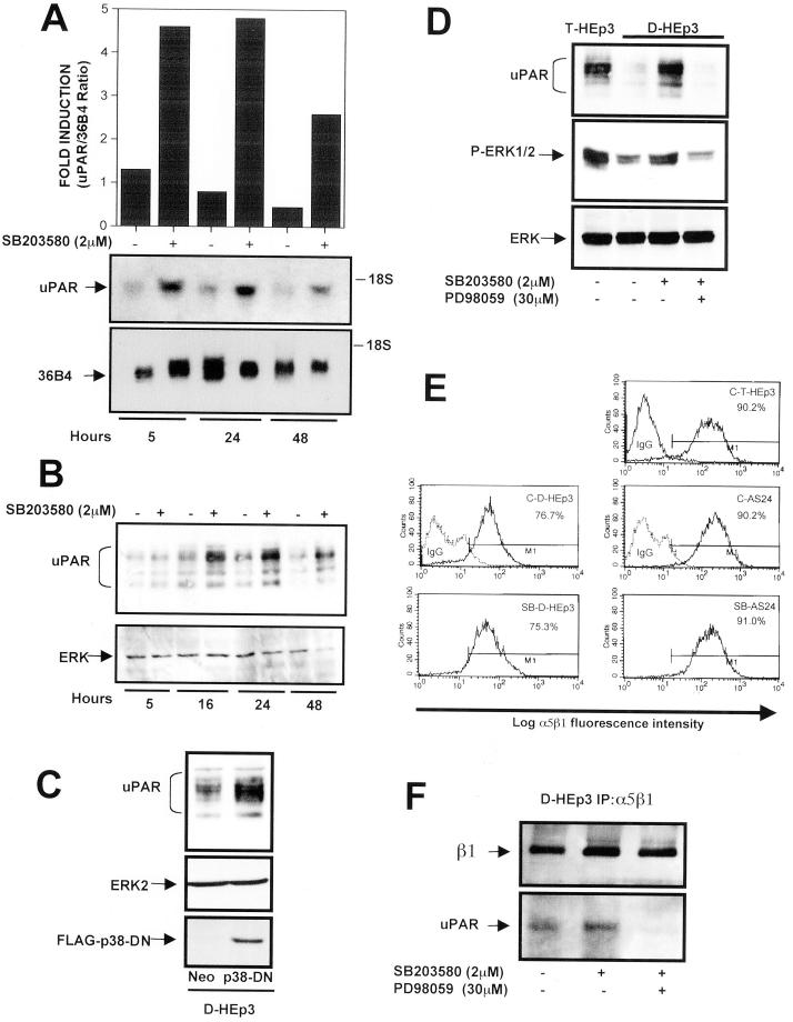
Figure 5.
Effect of p38 inhibition on uPAR and α5β1 expression (A) uPAR-mRNA level. RNA extracted from D-HEp3 cells treated with DMSO (−) or 2 μM SB203580 in DMSO (+) for 5, 24, and 48 h were tested by Northern blot using 32P-labeled uPAR-cDNA as a probe. Bottom, 36B4 mRNA used as a loading control. The graph shows the ratio of uPAR/36B4 signals, measured by laser scanning densitometry. (B) uPAR-protein level. D-HEp3 cells were incubated with 2 μM SB203580 in DMSO (+) or DMSO (−) for 5, 16, 24, or 48 h and, after lysis, the cells were processed and analyzed by Western blotting for uPAR. Total ERK served as a loading control. (C) uPAR protein levels in cells expressing a dominant negative p38. D-HEp3-neo or D-HEp3-p38DN cells were lysed and the levels of uPAR, ERK (as loading control), or p38DN were detected by Western blot. (D) Effect of Mek inhibition on SB203580-induced ERK activation and uPAR up-regulation. D-HEp3cells untreated, or treated with 2 μM SB203580 with or without 30 μM PD98059 (Mek inhibitor) for 36 h, were lysed, and the levels of uPAR protein (top) as well as active (middle) and total (bottom) ERK levels were detected by Western blot. The levels of uPAR and active ERK in T-HEp3 cells served as positive controls. (E) FACS analysis for α5β1-integrin surface expression using anti-α5β1-integrin antibodies (clone HA5) in T-HEp3, D-HEp3, or AS24 cells treated with the p38 inhibitor SB203580 (2 μM) for 48 h (SB-D-HEp3 or SB-AS24) or untreated (C-T-HEp3, C-D-HEp3, C-AS24). Isotype-matched IgG was used as control. The numbers indicate the percentages of cells positive for surface α5β1-integrin. (F) Coimmunoprecipitation of uPAR and α5β1-integrin in SB203580-treated cells. D-HEp3 cells untreated or treated with 2 μM SB203580 or 2 μM SB203580 and 30 μM PD98059 were lysed, and the cell lysates were subjected to IP with anti-α5β1-integrin antibodies and the immunoprecipitated proteins were tested by Western blotting with antibodies to β1-integrin and uPAR.

