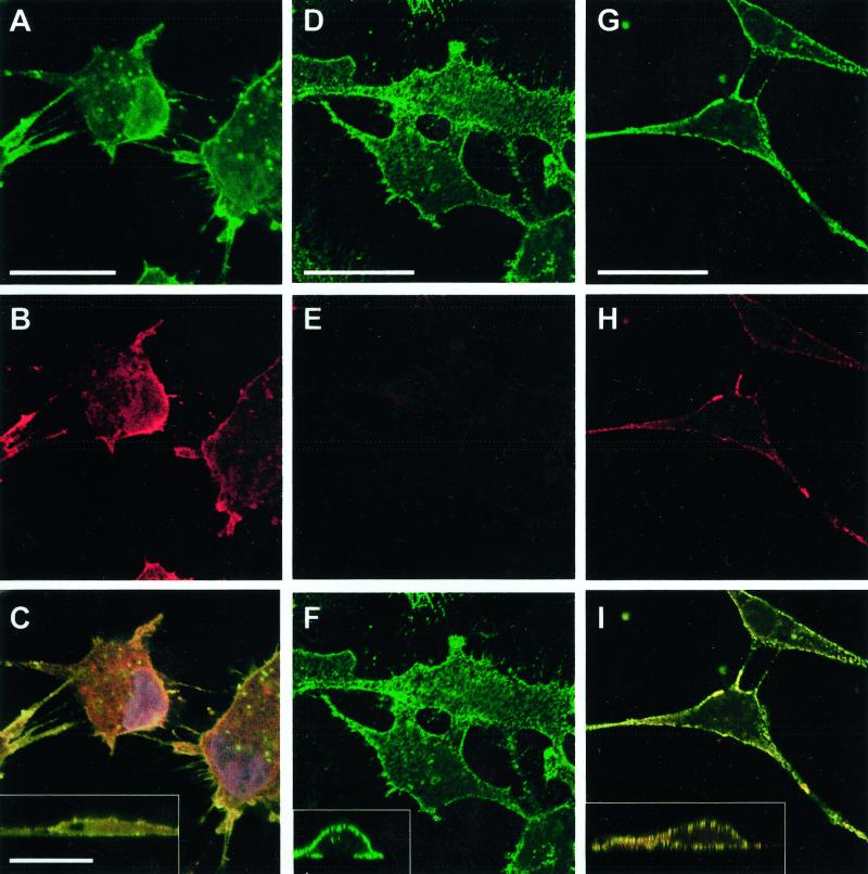
Figure 6.
Effect of p38 inhibition on uPAR and β1-integrin surface expression. Untreated T-HEp3 cells (A–C) or D-HEp3 cells treated for 48 h with 2 μM SB203580 (G–I) or DMSO alone (D–F) were fixed and stained, without permeabilization, for β1-integrin (A, D, and G, AIIB2 antibody, green) or uPAR (B, E, H, R2 antibody, red), as described in MATERIALS AND METHODS, and detected by confocal laser scanning IF microscopy. C, F, and I, show the merged image of the uPAR and β1-integrin signals; yellow indicates colocalization of uPAR and β1-integrin signals. A to I are XY sections; insets in C, F, and I are XZ sections of the same cells showing the overlay. Note the high surface (apical and basolateral) expression of uPAR in D-HEp3 cells treated with the p38 inhibitor (compare E with H) that colocalizes with β1-integrin signal (compare F with I and the corresponding insets), which remains unchanged in SB203580-treated cells (compare D and G). Bars, 40 μm.

