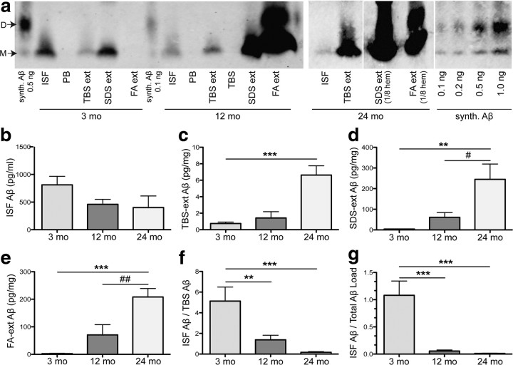
Figure 4.
Aβ in all pools of brain parenchyma accrue with age while those that remain diffusible in the ISF declines. a, Representative IP/WBs of Aβ species from brains of the same mice right after microdialysis, in four pools: ISF, TBS extracted (ext), SDS extracted, and FA extracted. All pools (except FA extracted, which was lyophilized and straight-loaded onto the gel) were immunoprecipitated with AW8 and blotted with 6E10 and 4G8. Synthetic (synth.) Aβ run alongside for quantification. Perfusion buffer (PB) and TBS were immunoprecipitated as negative controls. b–e, Quantification of IP/WBs from 21 mice shows ∼50% decrease in absolute values of ISF Aβ between 3 and 12 months (not significant by one-way ANOVA followed by Bonferroni test) (b), with a sharp rise in TBS- (c), SDS- (d), and FA-(e) extracted Aβ (picograms per milligrams wet brain tissue). f, g, Ratios of ISF to TBS-soluble Aβ (f) or to total parenchymal Aβ (g) calculated for each mouse and shown as mean ratio ± SEM; n = 7 mice per group. Aβ quantified by Licor Odyssey imaging and analyzed by one-way ANOVA and Bonferroni test: **p < 0.01 and ***p < 0.001 versus 3 months; #p < 0.05 and ##p < 0.01 versus 12 months. D, Dimers; M, monomers.

