
Figure 4.
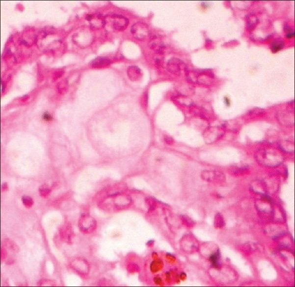
Tumor cells arranged in form of follicles containing eosinophillic colloid with scalloped edges (H and E stain, 400×)
Official websites use .gov
A
.gov website belongs to an official
government organization in the United States.
Secure .gov websites use HTTPS
A lock (
) or https:// means you've safely
connected to the .gov website. Share sensitive
information only on official, secure websites.
Tumor cells arranged in form of follicles containing eosinophillic colloid with scalloped edges (H and E stain, 400×)