Figure 9.

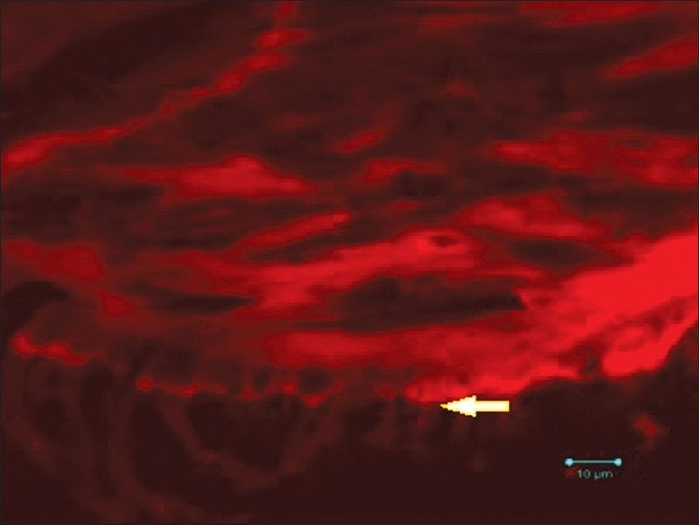
Confocal microscopic view (high power) of the psammoma bodies exhibiting varying densities of calcifications and brush border (Alizarin red, ×100 – Case 2)

Confocal microscopic view (high power) of the psammoma bodies exhibiting varying densities of calcifications and brush border (Alizarin red, ×100 – Case 2)