Figure 3.

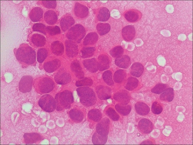
FNA smear showing a cluster of small round cells showing scanty cytoplasm, granular chromatin, absent nucleoli, and moderate anisonucleosis (H and E, 400×)

FNA smear showing a cluster of small round cells showing scanty cytoplasm, granular chromatin, absent nucleoli, and moderate anisonucleosis (H and E, 400×)