Figure 3.

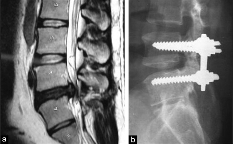
(a) T2W sagittal MRI showing L5-S1 prolapsed intervertebral disc. (b) X-ray of lumbar spine (lateral view) showing grade 3 fusion after instrumented laminectomy bone chip PLIF

(a) T2W sagittal MRI showing L5-S1 prolapsed intervertebral disc. (b) X-ray of lumbar spine (lateral view) showing grade 3 fusion after instrumented laminectomy bone chip PLIF