
Figure 9.
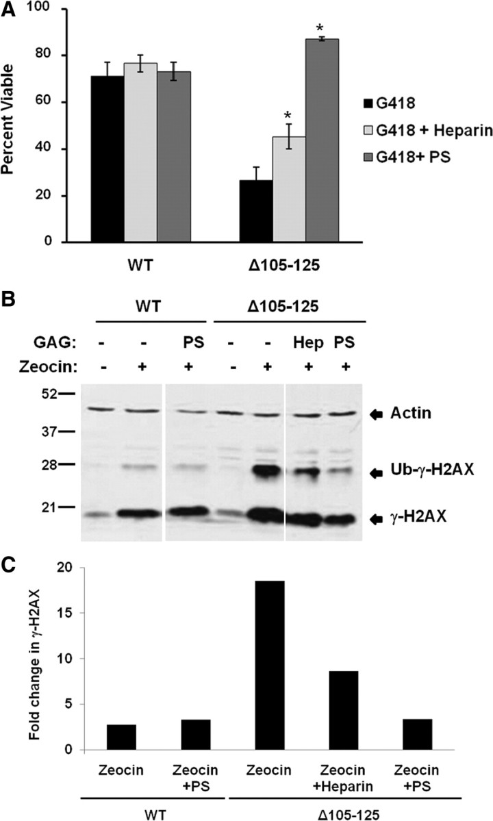
GAGs suppress the toxicity of Δ105–125 PrP in cell culture. A, HEK cells expressing WT or Δ105–125 PrP were treated for 48 h in the presence or absence of 400 μg/ml G418, after which cell viability was measured by MTT reduction. Cell viability is expressed as the value for MTT reduction (A570) of G418-treated cells as a percentage of the value for untreated cells. Where indicated, PS or heparin (each at 100 μg/ml) were present during the 48 h incubation period. The bars show mean values ± SEM. The asterisks indicate values that are significantly different between PS- or heparin-treated cells compared with cells not treated with GAGs (p < 0.0001). B, HEK cells expressing WT or Δ105–125 PrP were pretreated for 1 h in the presence of absence of heparin (“Hep”) (100 μg/ml) or PS (100 μg/ml), after which incubation was continued for an additional 30 min in the presence or absence of Zeocin (400 μg/ml). Cells were then lysed and analyzed for phosphorylated H2AX (γ-H2AX) and actin by Western blotting. The positions of γ-H2AX, ubiquitinated γ-H2AX (Ub-γ-H2AX), and actin are indicated. C, Western blot signals for γ-H2AX were quantitated and normalized to the amount of actin. γ-H2AX levels in the presence of Zeocin were expressed as a fold change relative to levels in the absence of Zeocin. A single experiment, representative of at least three similar ones, is shown.