Figure 1.
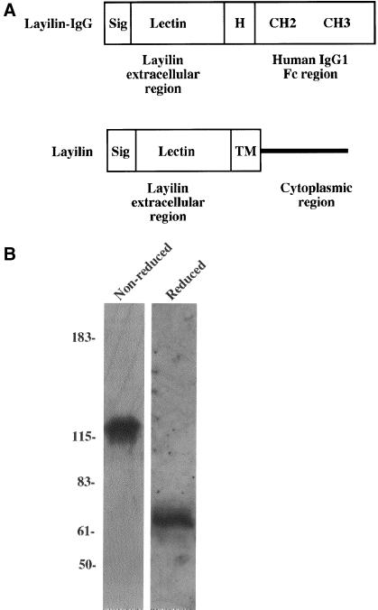
Structure and purification of recombinant layilin-Fc fusion protein. (A) The protein domains of the layilin-Fc and wild-type layilin are shown. Layilin's extracellular part was cloned immediately N-terminal to the hinge domain (H) of the human IgG1 so that the chimera contains two cysteine residues (not shown) within the hinge domain responsible for Ig dimerization. Sig, NH2-terminal signal sequence, Lectin, layilin's extracellular part, which is homologous with C-type lectins; TM, transmembrane domain; CH2 and CH3, constant regions of the human IgG. (B) Purified layilin-Fc fusion protein was analyzed on an SDS-PAGE gel under both reduced and nonreduced conditions and detected by immunoblotting with the anti-human IgG (Fc-specific) antibody. Molecular masses (in kDa) are shown at the left.

