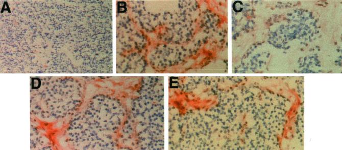
Figure 5.
Histochemical staining of tumors derived from pancreata of RIP-Tag2 mice. Cryostat sections of mouse pancreas tumors were reacted with chimeras (0.5 μg/ml) and processed for histochemistry as described in MATERIALS AND METHODS. Original magnification 50× in A and 400× in B–E. (A) Control fusion protein (E-cadherin IgG) staining of tumor section. The ECM lacks staining. (B) Layilin-IgG stains positively the ECM, and the staining is sensitive to hyaluronidase treatment before incubation with layilin-IgG (C). Similar pretreatment of sections with chondroitinase (D) or heparitinase (E) did not abolish layilin-Fc reactivity.

