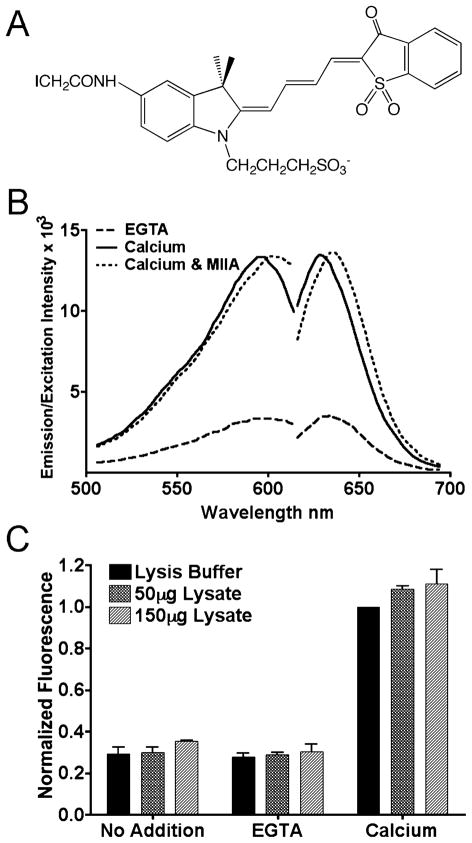
Figure 1.
Mero-S100A4 reports activation by Ca2+. (A) Structure of the I-SO merocyanine dye. (B) Fluorescence excitation and emission spectra of 5 μM Mero-S100A4 dimer. Dashed line: Mero-S100A4 in the presence of EGTA. Solid line: Mero-S100A4 in the presence of Ca2+. Dotted line: Mero-S100A4 in the presence of Ca2+ and myosin-IIA. Mero-S100A4 exhibits a 3-fold increase in fluorescence upon Ca2+ addition. The addition of Ca2+ and a 10-fold molar excess of myosin-IIA results in a slight red shift, but no additional increase in fluorescence intensity. (C) Mero-S100A4 was added to a fibroblast lysate in the presence of EGTA or calcium. The data represent the average peak intensity at 634 nm for three independent experiments and the standard deviation.

