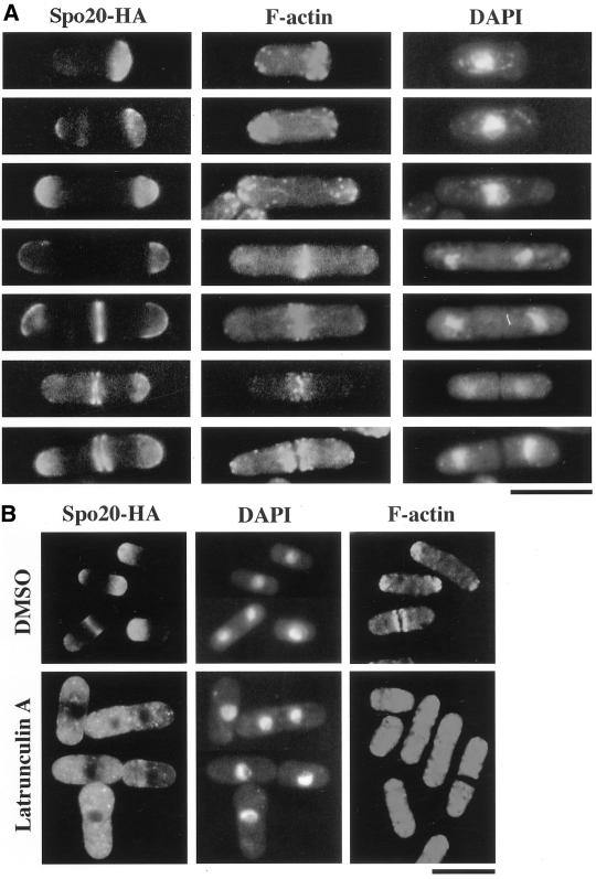
Figure 8.
The Spo20 localizes in close proximity to the F-actin cytoskeleton in vegetative cells. (A) Fluorescence microscopy using anti-HA and rhodamine-phalloidin. Wild-type strain YN8-WH carrying spo20-HA was grown to midlog phase in SSL at 28°C and then fixed. Cells were stained with anti-HA antibody to detect Spo20, with rhodamine-phalloidin to detect F-actin and with DAPI to visualize nuclear chromatin regions. (B) Disruption of the Spo20 localization by depolymerization of the F-actin cytoskeleton. Wild-type strain YN8-WH carrying spo20-HA was treated with latrunculin A for 2 h. Cells were processed for immunofluorescence microscopy to visualize F-actin and Spo20 and to stain nuclear chromatin regions. Bars, 10 μm.

