Figure 2.
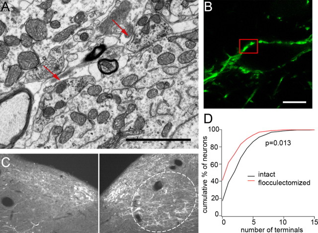
Purkinje cell terminals in the magnocellular MVN originate in the ipsilateral flocculus and form functional synapses. A, B, Ultrastructure of GFP-positive Purkinje cell terminals was visualized by scanning electron microscopy. A, Image of ultrastructure of Purkinje cell terminals surrounding a dFTN. Symmetrical synapses and numerous vesicles (arrows) in the bouton indicate that they form functional synapses. B, GFP-positive Purkinje cell terminals surrounding a dFTN. Boxed area was magnified for examining the ultrastructure of synapses shown in A. C, GFP-positive Purkinje cell terminals in the ipsilateral MVN were mostly removed after unilateral surgical ablation of the flocculus (left), while those on the contralateral side of the MVN were intact (dotted area, right). D, The numbers of neurons innervated by Purkinje cells were counted on the intact (contralateral) and flocculectomized (ipsilateral) side of the MVN and the data are represented as a cumulative graph. Note the increased numbers of non-FTNs after surgical ablation of the flocculus. Scale bars: A, 2 μm; B, 10 μm.

