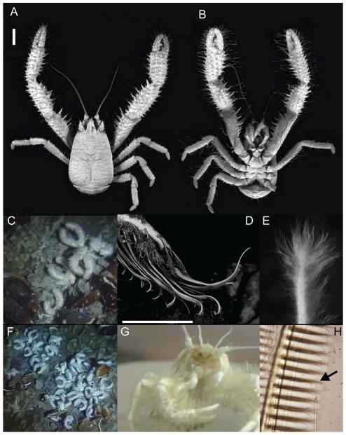
Figure 1. Kiwa puravida holotype, in situ images, and morphologic and behavioral adaptations to harvesting its bacteria.
Kiwa puravida male holotype: (A) Dorsal view. (B) Ventral view (A and B Scale bar = 10 mm; Credit: Shane Ahyong, NIWA Wellington). (C) in situ next to Bathymodiolin mussels (D) Scanning Electron Micrograph of a detail of K. puravida's 3rd maxilliped and the comb-row setae which it uses to harvest its bacteria (scale bar = 150 µm credit; Shana Goffredi, Occidental College]. (E) Setae covered by bacteria from 3rd pereopod (see Figure 4E for scale). (F) Dense aggregation in situ. (G) Shipboard photo of K. puravida using its 3rd maxilliped to harvest its epibiotic bacteria. (H) Comb-row setae with bacteria filaments stuck among combs (indicated by arrow).

